
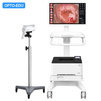
OPTO-EDU A43.1932 Digital Colposcope Continous Zoom 1-90x Gynecology
-
Highlight
Digital Colposcope 1-90x Zoom
,Gynecology Surgical Operating Microscope
,Continuous Zoom Colposcope
-
Magnification1-90x
-
Working DistanceAt 3X Magnification: 240mm-380mm
-
Filed Of View (FOV)At 3X Magnification: Dia. 60mm
-
Depth Of Field (DOF)At 4X Magnification: >40mm; At 18X Magnifcation: >5mm
-
Horizontal ResolutionSystem Imaging Horizontal Resolution >1350 TVL
-
Spatial ResolutionResolution At The Center Of FOV >20 Ip/mm
-
Color ReproductionMax Color Reproduction Error For A Single Color Patch
-
Geometric DistortionSystem Imaging Geometric Distortion ≤3%
-
Illumination9 LED Light, Multispectral Light Source Imaging
-
Spot DiameterSpot Diameter At 300mm From Light Source: 85mm
-
Place of OriginChina
-
Brand NameCNOEC, OPTO-EDU
-
CertificationCE,
-
Model NumberA43.1932
-
Document
-
Minimum Order Quantity1 pc
-
PriceFOB $1~1000, Depend on Order Quantity
-
Packaging DetailsCarton Packing, For Export Transportation
-
Delivery Time5~20 Days
-
Payment TermsT/T,West Union,Paypal
-
Supply Ability5000 pcs/ Month
OPTO-EDU A43.1932 Digital Colposcope Continous Zoom 1-90x Gynecology
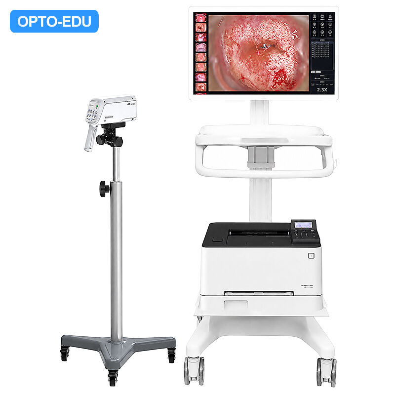
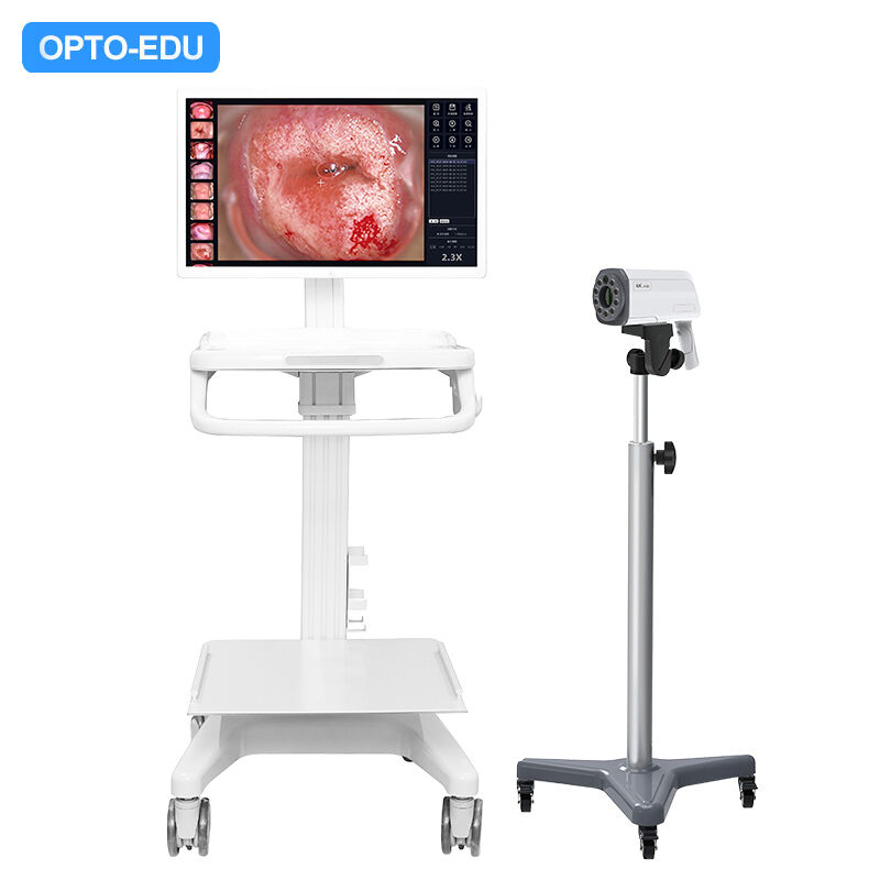
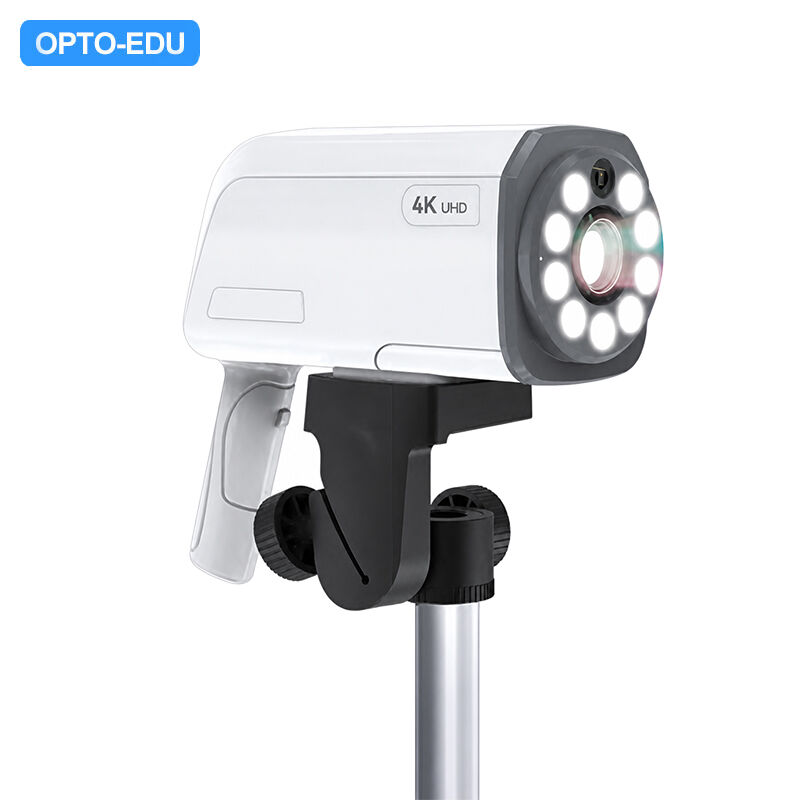
The OPTO-EDU A43.1932 is a professional digital video colposcope engineered for gynecological examination and diagnosis. Built around a Sony Exmor CMOS sensor and a continuous 1x–90x zoom optical system, it delivers sharp, true-to-life cervical imaging with a working distance of 240–380 mm and an auto measurement range of 180–380 mm. The integrated control panel provides manual and auto focus, manual and auto white balance, zoom, freeze, and timing functions, while HDMI output connects directly to a 24-inch PC workstation supplied with professional colposcopy software and a laser printer.
- Digital camera: Sony Exmor CMOS sensor, resolution 1920×1080P or 3840×2160P (4K model)
- Total magnification: 1x–90x continuous zoom, working distance 240–380 mm, auto measure range 180–380 mm
- Control panel: Manual/auto focus, manual/auto white balance, zoom, freeze, and timing
- Output & workstation: HDMI output to 24" PC workstation with professional software and laser printer
- Illumination: Multispectral LED light source, >7500 LUX, built-in 3-level green and white filter

Powered by the Sony 4K EXMOR CMOS image sensor, the new-generation 4K UHD video colposcope delivers exquisitely sharp imagery with rich, true-to-life colors, rendering three-dimensional and highly detailed UHD images. Integrated with colposcopy management software, it provides clinicians with reliable support for precise examination and diagnosis.
| Items | Digital Video Colposcope | A43.1932 | A43.1932-4K |
|---|---|---|---|
| Magnification | 1–90x | ● | ● |
| Sensor | Sony EXMOR CMOS | ● | |
| Sensor (4K) | Sony 4K EXMOR CMOS | ● | |
| Imaging Resolution | Max image capture resolution up to 1920×1080 | ● | |
| Imaging Resolution (4K) | Max image capture resolution up to 4K 3840×2160 | ● | |
| Working Distance | At 3X magnification: 240 mm–380 mm; at max magnification: 260 mm–300 mm | ● | ● |
| Field of View (FOV) | At 3X magnification: Dia. 60 mm; at max magnification: >Dia. 4 mm | ● | ● |
| Depth of Field (DOF) | At 4X magnification: >40 mm; at 18X magnification: >5 mm | ● | ● |
| Horizontal Resolution | System imaging horizontal resolution >1350 TVL | ● | ● |
| Spatial Resolution | Resolution at the center of FOV >20 lp/mm | ● | ● |
| Color Reproduction | Max color reproduction error for a single color patch on ColorChecker chart 30 NBS; average error 20 NBS; average camera saturation 93%–120% | ● | ● |
| Geometric Distortion | System imaging geometric distortion ≤3% | ● | ● |
| Illumination | 9 LED light, multispectral light source imaging; at 200 mm working distance >15,000 lux; at 300 mm working distance >7,500 lux | ● | ● |
| Spot Diameter | Spot diameter at 300 mm from light source: 85 mm | ● | ● |
| Color Temperature | 4000K–6500K | ● | ● |
| Color Rendering Index (CRI) | Ra ≥92 | ● | ● |
| Light Source Temp. Rise | Temperature rise on the illuminated surface after 20 mins <2°C | ● | ● |
| Radiant Heat | Max radiant intensity within FOV at 250 mm working distance ≤350 W/m² | ● | ● |
| UV Irradiance | Max effective UV irradiance within 200 nm–400 nm spectral range ≤5 W/m² | ● | ● |
| Light Source Uniformity | Max illuminance / average illuminance <1.5 within specified illumination range (spot diameter) | ● | ● |
| Auto Distance Measurement | Measurement accuracy error within 180 mm–380 mm range ≤15% | ● | ● |
| Signal Output | HDMI output to monitor through TMDS protocol | ● | ● |
| Control Panel | Auto & manual focus, manual zoom-in and zoom-out; 3-level white light and 3-level green light electronic filter imaging; auto & manual white balance adjustment; automatic timing function for the acetic acid test with customizable duration (timing mark displayed on live preview, captured images, and printed reports); magnification display on live preview, captured images, and printed reports | ● | ● |
| Software Measurement Accuracy | Software provides length and area measurement functions with an accuracy error of 5% | ● | ● |
| Stand System | Adjustable height range: min height <900 mm, adjustable range >190 mm; pan/tilt pitch angle >30° | ● | ● |
| Image Processor | All-in-one workstation, 7th Gen Intel Core i5, RAM 4 GB, storage 256 GB, 23.8-inch high-brightness LCD monitor, with relevant software and drivers | ● | ● |
| Report Output | Color laser printer | ● | ● |
| Operating Environment | Ambient temperature 5°C–35°C; relative humidity <80%; atmospheric pressure 86 kPa–106 kPa | ● | ● |
| Storage & Transport Environment | Ambient temperature -40°C–+55°C; relative humidity <80%; atmospheric pressure 86 kPa–106 kPa | ● | ● |
| Operating Voltage | ~220V±22V, 50Hz±1Hz | ● | ● |
Powered by multispectral imaging technology, the system integrates white light and narrow-band illumination to enhance vascular visualization. By leveraging hemoglobin's absorption of blue and green light, it improves the visibility of cervical vascular patterns, micro-lesions, and lesion boundaries for more detailed clinical observation.
The ergonomic multi-functional control panel features high-performance all-in-one shortcut keys for quick adjustment of magnification and focus, one-touch laser distance measurement, and convenient one-handed operation. The integrated upright mobile imaging stand with joystick control ensures stable positioning during procedures, enhancing workflow efficiency and user experience.
Our products are sold all over the world, you can rest assured.