
Opto Edu A64.0960 Laser Confocal Scanning Microscope Full Auto Motorized
-
Highlight
A64.0960 Laser Confocal Scanning Microscope
,Laser Confocal Scanning Microscope
,Full Auto Motorized Scanning Microscope
-
Scan AreaXY Scanning System
-
Resolution512×512~4096×4096
-
Pixel0.5us~8us
-
Zoom Scanning1-16X
-
FL Detector 3pcs3PMT
-
NosepieceMotorized, 6-holes
-
Place of OriginChina
-
Brand NameCNOEC, OPTO-EDU
-
CertificationCE, Rohs
-
Model NumberA64.0960
-
Document
-
Minimum Order Quantity1 pc
-
PriceFOB $1~1000, Depend on Order Quantity
-
Packaging DetailsCarton Packing, For Export Transportation
-
Delivery Time5~20 Days
-
Payment TermsT/T, West Union, Paypal
-
Supply Ability5000 pcs/ Month
Opto Edu A64.0960 Laser Confocal Scanning Microscope Full Auto Motorized
|
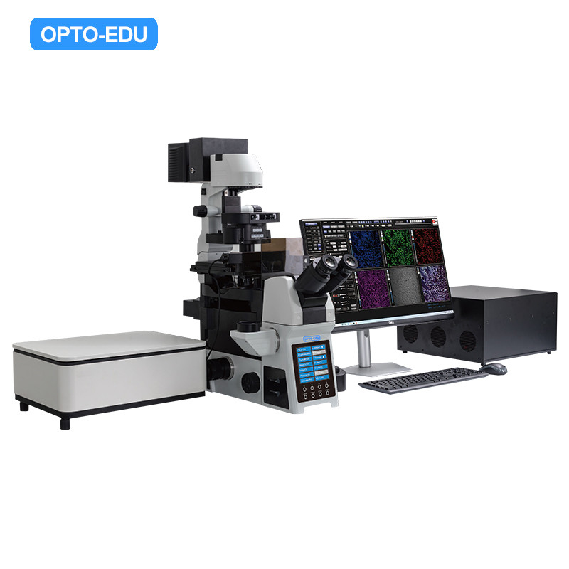
▶ Laser Light 405nm/50mW, 488nm/50mW, 561nm/50mW, 640nm/40mW
▶ Confocal Scan Module X/Y dual axis high speed optical scanning, resolution 4Kx4K, field of view 19mm, zoom scanning 1x-16x
▶ Probe Unit Standard 3 MA PMTs, GaAsP PMT Optional
▶ Microscope Inverted Fluorescent Microscope, full motorized, infinity plan SUPER APO objective 10x20x40x60x100x
▶ Camera 5 million pixels, color camera, SONY IMX264, frame rate 35fps, USB3.0, 0.65x C-Mount
▶ Software Photo and preview parameter self-adaptive; two rescan processing; image rotation output; full vision and ROI scanning image, support XY, XYZ, XYZT
▶ Computer CPU: Intel I7 or above, RAM: ≥16G, Hard disc: ≥1T+256G, Graphics card: Single, Monitor: ≥27 inch, resolution 2560×1440, Operation system: 64 bite, Windows X10 |
|
▶ Innovative Confocal Pinhole Unit Pinhole design is based on the principle of light reversibility. The excitation light of the lamp and the emission light of the sample pass through the same pinhole, and they keep a 100% conjugate relationship. It not only ensures the acquisition efficiency of fluorescence signal, but also improves the filtering of non-focal plane signal, for the higher detection sensitivity and better image resolution. |
|
▶ Controller Probe Unit The probe unit consists of a 6-position electric filter turntable with 4 filters as standard and a single high-sensitivity multi base photomultiplier tube (MA PMT, QE≥25%@500nm), is able to easily and quickly automatically complete multicolor fluor escence confocal imaging. |
|
|
▶ APO Series Apochromatic Objectives Converging the optic axises of red, green and blue to one focal plane, correcting the axial chromatic aberration of violet light, the original color of samples is able to be presented. And the resolution and effective magnification are improved based on large numerical aperture. |
|
▶ SAPO Series SUPER Apochromatic Objectives With large numerical aperture, excellent color difference correction and flat field, more uniform, bright and high- resolution fluorescence images can be obtained. |
|
▶ Cell Imaging A64.0960 is able to accurately image all cells labeled with various fluorescent proteins and multicolor probes, studying the fluorescence colocalization, dynamic properties and spatial relationships of two or more target proteins. Besides, A64.0960 can achieve the morphological structure of 3D cell culture such as organoids/globules by 3D reconstruction, finding out more hidden information. |
|
▶ Histopathological sections of animals and plants The layer scanning of A64.0960 is suitable for different histopathological sections of animals and plants, especially for large tissue. Much more details and more accurate data are available. |
|
▶ Confocal Software Support single channel or multi-channel 2D Imaging (XY), 3D imaging (XYZ), 4D imaging (XYZT) and multi-site scanning. It is available for imaging, photobleaching and photo stimulation within a user-defined ROI, as well as Z- Stack imaging, jigsaw puzzles, scale correction, filtering processing, data recording, etc. |
| A64.0960 Laser Confocal Scanning Microscope, Full Auto Motorized | |||
| Item | Specification | Qty | |
| A64.0960 | Laser Confocal Scanning Microscope, Full Auto Motorized, Standard Outfit | ||
| Main Body | Laboratory Level Inverted Microscope, Including: --Viewing Head, Main Body, --LED Transmit Illumination System, --L Type Fluorescent Reflect Illumination System, --Motorized Fluorescent Disc --Motorized 7 Holes Universal Turret Disc Condenser For BF, DF, FL, DIC. --Motorized Controller, --Power Cord, Data Cables, --Fluorescent Oil |
● | |
| View Head | 20-45° Inclined, Gemel Binocular, Interpupillary 50~76mm | ● | |
| Eyepiece | PL10X/22mm, Diopter Adjustable | ● | |
| Nosepiece | Motorized 6 Holes Nosepiece | ● | |
| Motorized Stage | Motorized Mechanical 3 Layers Working Stage, --Size 350mm(X) x 200mm(Y), Moving Range 114mm(X) x 75mm(Y), --Absolute Positioning Accuracy <2um/10mm --Unidirectional Repeat Positioning Accuracy <1um, --Bidirectional Repeat Positioning Accuracy <2.5um, --Maximum Speed 50mm/s, --Including Motorized Contoller, Slide Holder T-SGH1 |
● | A54.0964 |
| Ø36 Petri Dish Holder | ● | A54.0964-36 | |
| 96 Holes Dish Holder | ○ | A54.0964-96 | |
| Terasaki Holder | ○ | A54.0964-T | |
| Infinity Plan Apochromatic Objective: |
Infinity Plan APO 4X/0.16 WD=12.8mm | ○ | A5F.0962 |
| Infinity Plan Super-APO 10X/0.4 WD=3.1mm | ● | A5F.0963 | |
| Infinity Plan Super-APO 20X/0.8 WD=0.6mm | ● | A5F.0963 | |
| Infinity Plan Super-APO 40X/0.95 WD=0.18mm | ● | A5F.0963 | |
| Infinity Plan Super-APO 60X/1.42 WD=0.17mm, Oil | ● | A5F.0963 | |
| Infinity Plan Super-APO 100X/1.45 WD=0.14mm, Oil | ● | A5F.0963 | |
| Condenser: | Motorized, 7 Holes, N.A.>0.55, W.D.>27mm, 3 Holes For 30mm PH, 4 Holes For 38mm DIC, With Motorized Diaphragm, With Polarizer | ● | |
| Flourescent | 100W Mercury Fluorescent Illuminator, With Power Supply Box, With 100W Mercury Lamp (OSRAM) | ● | A5F.0960-WER |
| 5 Holes Motorized Filter Disc, With BF, Stopper, ND6/ND25/ND50 Filters, Including Cables | ● | ||
| 10W LED Fluorescent Illuminator, With 4 Channels: 365/460/525/625nm, Excite Controller Time <500ms, Brightness Separately Adjustable, Life Time >20000 Hours, Support SDK Control | ○ | A5F.0960-LED4 | |
| Extended Parts | ○ | A5F.0960-EX | |
| Fluorescent Filter B1,EX: AT480/30X, BS: AT505DC, EM:AT535/40M | ● | A5F.0960-B1 | |
| Fluorescent Filter G1,EX: AT560/40X, BS: AT600DC, EM:AT635/60M | ● | A5F.0960-G1 | |
| Fluorescent Filter UV1,EX: AT375/28X, BS: AT415DC, EM:AT460/50M | ● | A5F.0960-UV1 | |
| Fluorescent Filter R1,EX: AT620/50X, BS: AT655DC, EM:AT690/50M | ○ | A5F.0960-R1 | |
| DIC | DIC Imaging Kit For 10X, 20X, 40X, 60X, Including 1 DIC Detector, With 10-60X Differential Interference Imaging Function, Can Realize DIC-fluorescence Imaging And Overlay Analysis, Including DIC Insert Board, Analyzer Group, DIC Ring Plate | ● | |
| Laser Confocal | Laser Confocal XY Scan & Detector, 1 GaAsP | ● | AF.0965-GaAsP |
| Laser Confocal XY Scan & Detector, 1 PMT | ○ | AF.0965-PMT | |
| Semicoductor/Solid Laser Source With 4 Channels, Wavelength 405nm, 488nm, 561nm, 640nm, Output Power >50mW | ● | AF.0965-L1 | |
| Advanced Version Semicoductor/Solid Laser Source With 4 Channels, Wavelength 405nm, 488nm, 561nm, 640nm, Output Power >50mW | ○ | AF.0965-L2 | |
| Computer | HP i7-14700F RTX4060T1 Graphic Card, 32G Memory, 1TB HDD, Curved Monitor S3423DWC USB-C | ● | |
| Software | According To Different Imaging Modes, Multi-channel Time-division And Simultaneous Image Acquisition Can Be Achieved, Supporting Multiple Automatic Acquisition Processes Such as XY, XYZ, XYT, XYZT, etc., And Saving The Shooting Environment. Also Supports Large Image Stitching, 3D Reconstruction And Display, Cell Counting. With Dongle | ● | |
| Cable | Control Cable, USB-CAN Adapter | ● | |
| Platform | Professional Optical Shock Resistant Platform, Tabletop Size > 1000x800mm, Made of High Magnetic Conductivity Stainless Steel 1Cr17 Material | ○ | A54.0968 |
| Photo Port | Left Side Photo Port, Light Split 100:0 /0:100, Field of View 16mm, With Built-in 1x C-Mount | ● | A55.0960 |
| Right Side Photo Port, Light Split 100:0 /0:100, Field of View 16mm, With Built-in 1x C-Mount | ○ | A55.0960-R | |
| Camera | 5.0M Color 2/3” CMOS SONY IMX264, 35fps, USB3.0 | ● | A59.0960-5.0M |
| 20M Color 1.1” CMOS SONY IMX541, 17.5fps, USB3.0 | ○ | A59.0960-20MC | |
| 20M Monocolor 1.1” CMOS SONY IMX541, 17.5fps, USB3.0 | ○ | A59.0960-20MM | |
| Adapter | 0.65x C-Mount, Focus Adjustable | ○ | A55.0930-65 |
| 1.0x C-Mount, Focus Adjustable | ○ | A55.0930-10 | |
| Package | Strong Master Carton | ● | |
Our products are sold all over the world, you can rest assured.