
OPTO EDU A64.1020 Full Auto APO BF Laser Confocal Microscope with High-Speed Resonant Scanning and Spectral Imaging
-
Highlight
Full Auto Laser Confocal Microscope
,APO Laser Confocal Microscope
,BF Laser Confocal Microscope
-
EyepieceWF10x/22mm Eyepiece, Diopter Adjustable +/-5°, High Eyepoint
-
Head10-40° Tilt Adjustable Binocular Head
-
Mag. SwitchCoded Manual Intermediate Magnification Switch Button 1x/1.5x On Right Side
-
ObjectiveNIR Infinity Plan APO Achromatic Objective
-
FocusingMotorized Z Axis, Grating Type, Moving Range Up 8.5mm
-
Transmit LightKohler Illumination, With Field/Iris Diaphragm, 0~25° Titl Adjustable Arm
-
Place of OriginChina
-
Brand NameOPTO-EDU
-
CertificationCE, Rohs
-
Model NumberA64.1020
-
Document
-
Minimum Order Quantity1 pc
-
PriceFOB $1~1000, Depend on Order Quantity
-
Packaging DetailsCarton Packing, For Export Transportation
-
Delivery Time5~20 Days
-
Payment TermsT/T,West Union,Paypal
-
Supply Ability5000 pcs/ Month
OPTO EDU A64.1020 Full Auto APO BF Laser Confocal Microscope with High-Speed Resonant Scanning and Spectral Imaging
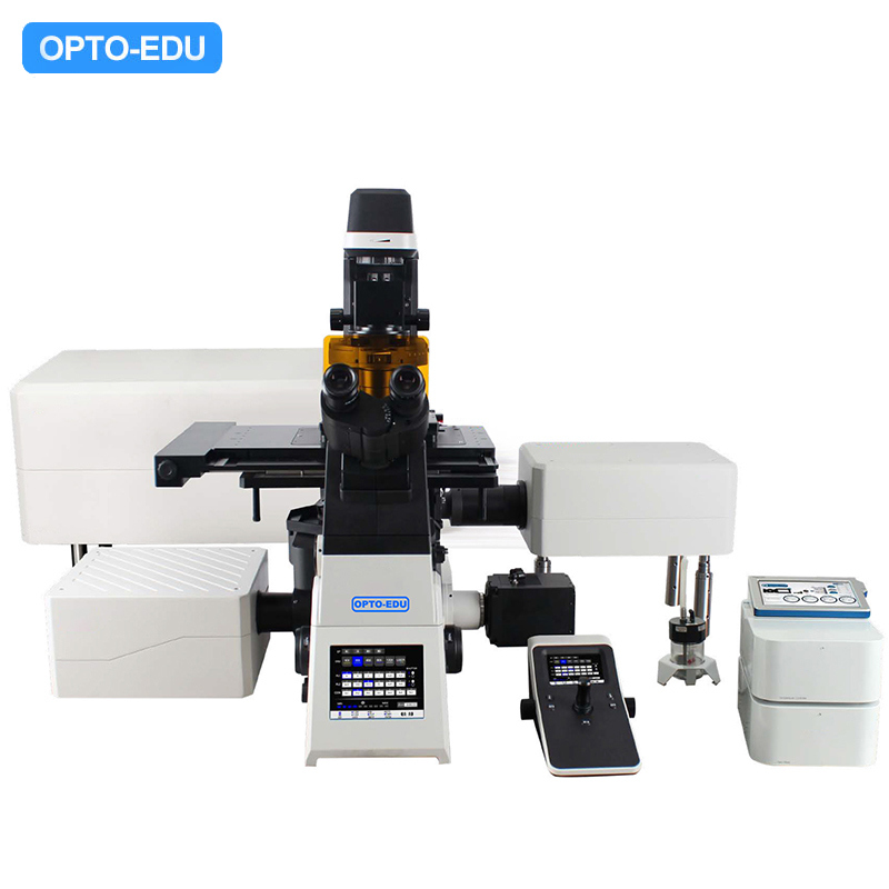
OPTO EDU A64.1020 NIR Laser Confocal Microscope Full Auto APO BF PH PL FL DIC Hoffman
The A64.1020 laser confocal microscope represents a significant advancement in biological research imaging technology. Designed for high-throughput, high-quality imaging, this system combines resonance scanning, array detection, and spectral imaging capabilities to meet the demanding requirements of modern life science research.
Key Features and Benefits
High-Speed Resonant Scanning
The A64.1020 utilizes resonant scanning technology, increasing scanning speeds by nearly 20 times while maintaining image quality. This reduces photobleaching and phototoxicity, enabling prolonged observation of sensitive live cell samples.
Gentle Live Cell Imaging
Reduced pixel dwell time minimizes laser exposure, extending cell viability while enabling high-frequency data acquisition. Ideal for drug development research and microplate screening applications.
Low-Noise, High-Resolution Detection
The array detector accurately captures weak fluorescence signals, delivering clear, detailed images with precise resolution of sample microstructures.
High Sensitivity Spectral Imaging
GaAsP photomultiplier tubes improve fluorescence acquisition efficiency, enabling observation of weak fluorescent samples. The system supports four spectral ranges with ≤ 2nm resolution for superior multicolor imaging.
Advanced Imaging Technologies
Space Array Detection
The SPAD detector array collects two-dimensional spatial information at each scan point, revealing ultrafine structures lost in traditional confocal detection.
Spectral Unmixing System
Precisely distinguishes substances with similar fluorescence characteristics through spectral analysis, enabling qualitative sample analysis and biomolecule identification.
Adaptive Focus System (AFS)
The intelligent AFS eliminates focus drift, delivering consistently sharp images across all imaging modalities including super-resolution and confocal imaging.
Precision Control and Operation
High-Speed Motorized Components
Rapid operation of objectives, filter cubes, XY stages, and observation modules creates an effortless workflow. The intuitive joystick control makes operation as natural as manual microscopy.
Multi-Dimensional Imaging
Supports combined X, Y, Z, λ, and T scanning with flexible modes including multi-channel fluorescence, time-lapse, Z-stacking, and panoramic stitching for diverse experimental needs.
Deconvolution Technology
Eliminates out-of-focus noise in confocal images through iterative deconvolution. 3D deconvolution enhances clarity and resolution across multiple axes for superior image quality.
Technical Specifications
| Component | Specification | Standard | Optional |
|---|---|---|---|
| Confocal System | 5 Channels Laser Confocal 400~750nm | ● | |
| Laser Source | 4 Laser Source With AOTF, Max Power 50mW (405nm, 488nm, 561nm, 640nm) | ● | |
| NIR Laser Source | 730nm | o | |
| Standard Detector | Wavelength 400-750nm, 5 Channels PMT (405nm, 488nm, 561nm, 640nm) | ● | |
| Spectral Detector | 4 Channels PMT, Spectral Range 400-750nm, Adjustable Accuracy 1nm | ● | |
| Array Detector | Array-based Single-pixel Photon Counter; Resolution 120nm | ● | |
| Scan Head | Max Pixel 8192x8192 (8K x 8K) | ● | |
| Computer System | i7-11700/32GB DDR4/1TB SSD/RTX A2000 6G/Win 11 | ● | |
| Monitor | 31" 4K Display | ● |
The A64.1020 offers exceptional expandability, supporting various accessories including live cell culture systems, super-resolution modules, and FRAP modules. This versatile system provides comprehensive solutions for cell biology, molecular biology, and biomedical research applications.
Related Products
Our products are sold all over the world, you can rest assured.